INTERACTIVE 3D MODEL OF THE SKIN
For more information on how to navigate the model, click on the question mark icon on the bottom right corner of the viewing screen.
EPITHELIUM OF THE SKIN
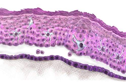
The epidermis of the skin is an epithelial tissue layer that covers the entire body, forming a barrier between the human body and the environment. Skin must be tough and therefore an epithelium with many layers that can be eroded and abraded without damaging the basement membrane is ideal. As such, skin consists of stratified squamous epithelium. For even tougher external surfaces a thick layer of keratin is produced on top of the squamous epithelium. Occasionally epithelium can change from one type to another (metaplasia) in response to injury. As it is a tough epithelium many tissues alter their surface to bear squamous epithelium.
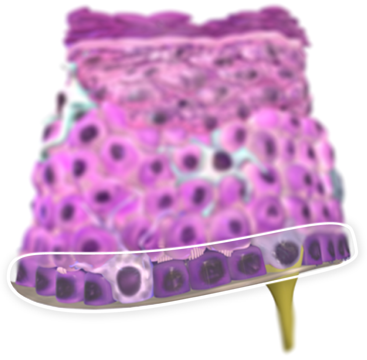
FIRST LAYER OF THE EPIDERMIS: THE BASAL LAYER
The deepest or basal epidermal layer is the germinal or proliferative cell layer.
This layer comprises of a single row of cells resting on a basal lamina, which is strongly adherent to the underlying dermis. The basal layer contains the replicative pool of germ cells. These cells give rise to daughter cells that continue to mature through the height of the epithelium.
This can be seen morphologically as basal cells have relatively little cytoplasm and appear much darker whereas cells in the upper layer have much more cytoplasm and appear much lighter.
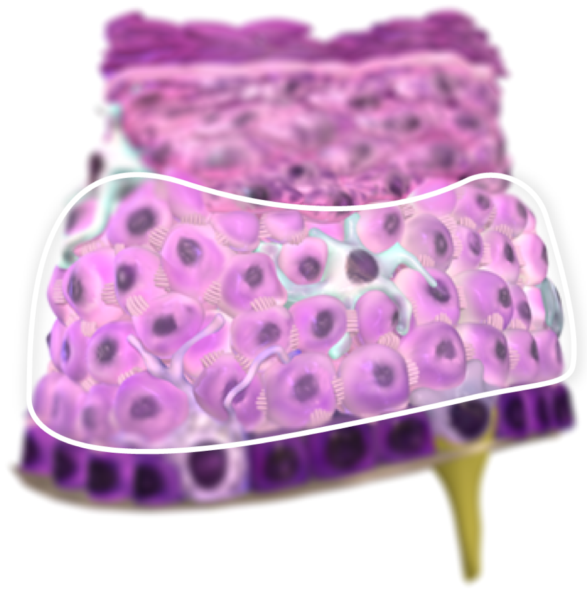
SECOND LAYER OF THE EPIDERMIS: THE PRICKLE CELL LAYER
The prickle cell layer represents the first area of maturation above the basal cells.
This layer is about five cells thick and is so named because the cells appear to have spines or prickles projecting from their surface. These are desmosomes on fine spike-like cytoplasmic processes; they interdigitate and attach to neighbouring cells.
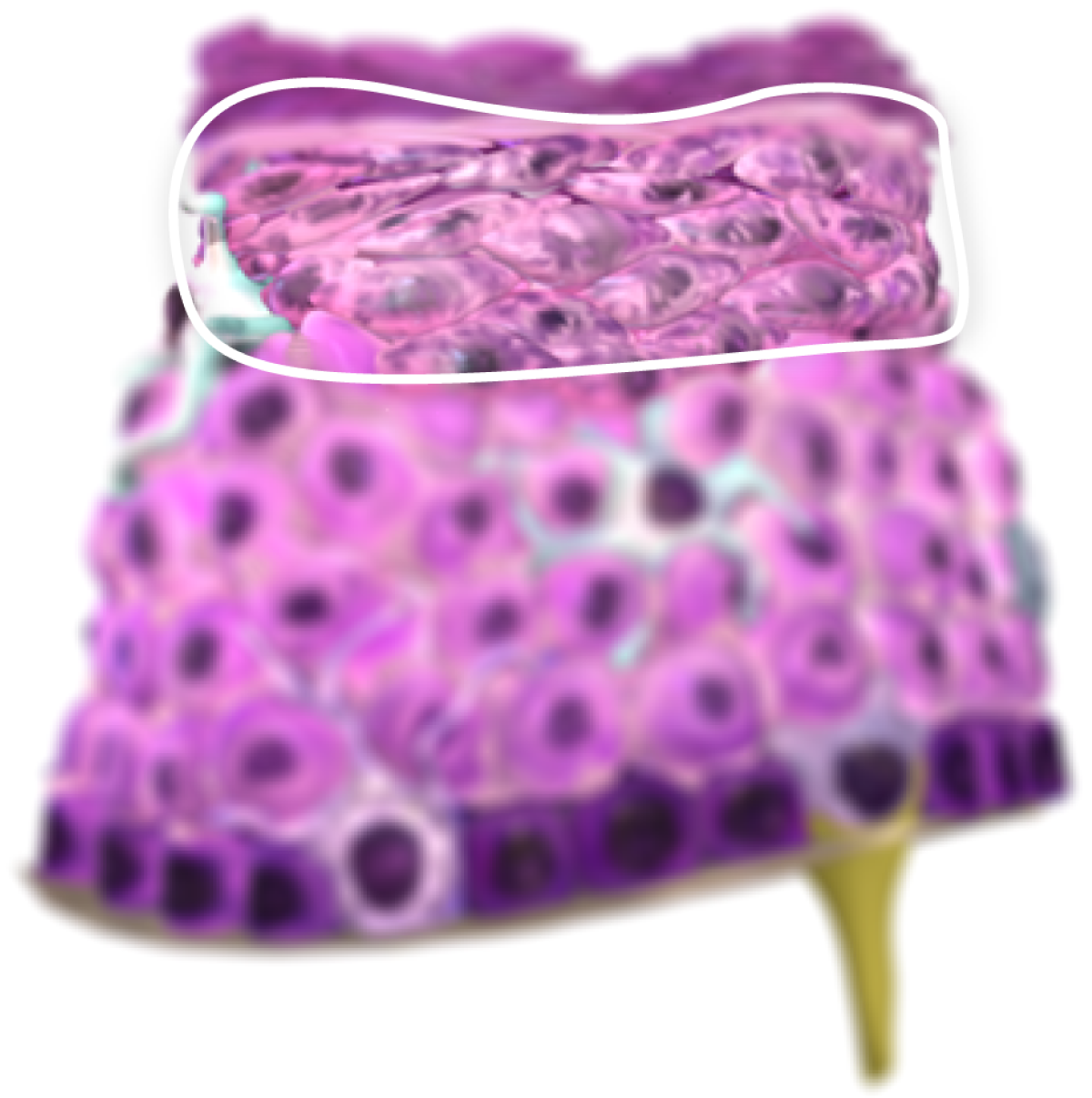
THIRD LAYER OF THE EPIDERMIS: THE GRANULAR LAYER
Transformation of the spinous cells into the cells of the third layer, the granular layer, is characterised by accumulation of numerous dense cytoplasmic granules containing proteins that eventually go on to form keratin. At the same time the nucleus and organelles break down and their ultimate destruction results in cells filled only with keratin. The cells of the granular layer have the distinction of being programmed to destroy their nuclei and organelles, yet at the same time to synthesize keratin and lamellar bodies.
The contents of the lamellar granules in the granular cells are discharged into the extracellular space and provide a lipid layer between the succeeding cell layers. This is effective in establishing permeability for the skin.
FOURTH LAYER OF THE EPIDERMIS: STRATUM LUCIDUM
In thick skin a narrow fourth layer, the stratum lucidum, is sometimes observed above the granular layer. It consists of flattened, dead cells with abundant keratin proteins and it is observed as a thin, undulating line of poor staining intensity.

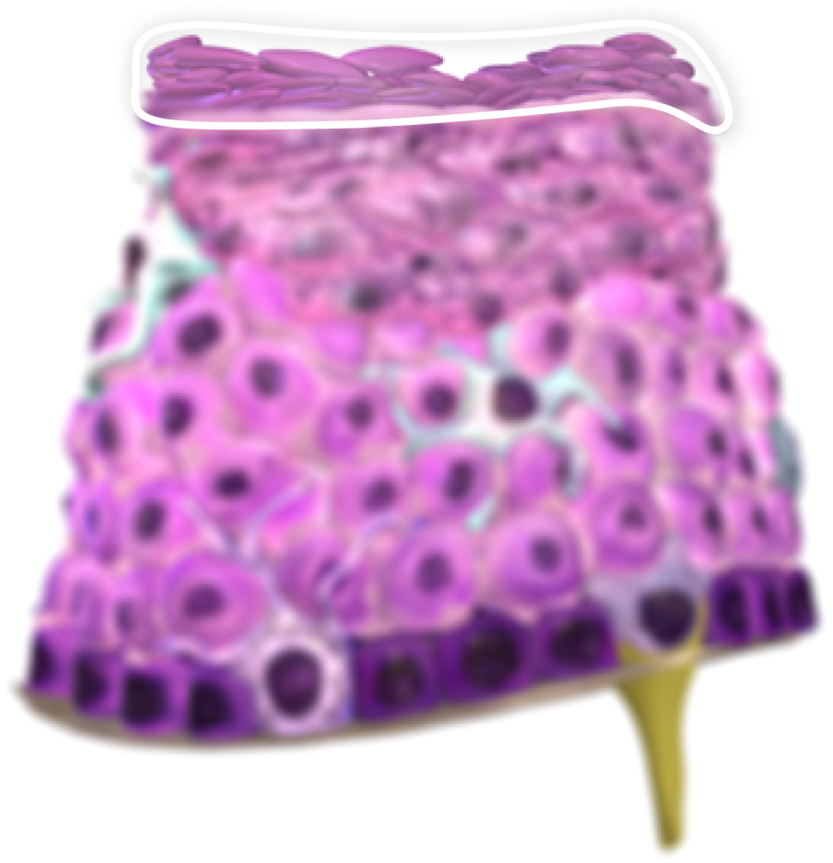
FIFTH LAYER OF THE EPIDERMIS: THE KERATIN LAYER
The fifth and most superficial layer is the keratin layer, which consists of dead, anucleate squamous cells containing keratin. it is especially thick (more than 1 mm) on the soles of the feet and quite thin (about 0.1 mm) over much of the body surface. Thus, the number of individual cell layers making up the fifth layer can range from about 10 to a few hundred. These plates of keratin, which are also referred to as the horny layer of cornified cells, are constantly shed from the surface and replaced by new cells arising from the deeper layers. Normally the transit time from a stem cell to desquamation is about 1 month.
CELLS RESIDENT IN THE EPIDERMIS: MELANOCYTES
Melanocytes synthesize melanin pigment from precursors such as tyrosine and dihydroxyphenylalanine (dopa) and transfer the pigment to surrounding keratinocytes within granules called melanosomes. Melanin absorbs and scatters the ultraviolet radiation that is present in sunlight and thereby protects cells from the possible mutagenic effects of ultraviolet light.
Differences in skin colour are related to the amount of melanin produced, since the number of melanocytes is similar in light and dark skin. Melanin production is increased in response to prolonged solar radiation, causing a suntan, whereas lack of melanin in albino conditions is associated with greater risk of epidermal damage and skin cancer.
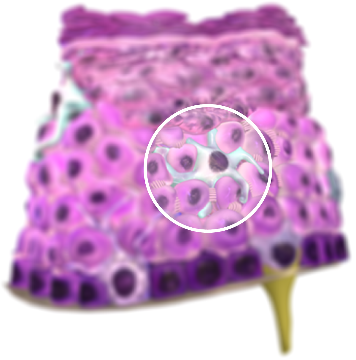
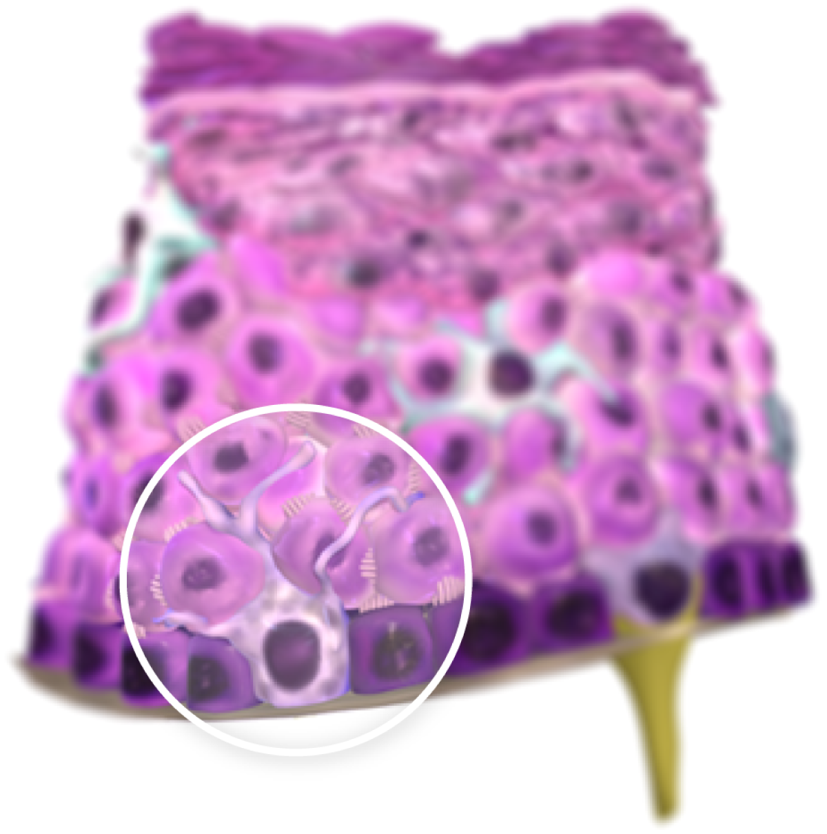
CELLS RESIDENT IN THE EPIDERMIS: LANGERHANS CELLS
The immunologic defence of the skin is in part attributed to Langerhans cells, which are found amongst the cells of the stratum spinosum. These cells migrate into the epidermis from the bloodstream; they function as antigen-presenting cells and may stimulate T cell responses in various allergic and inflammatory conditions.
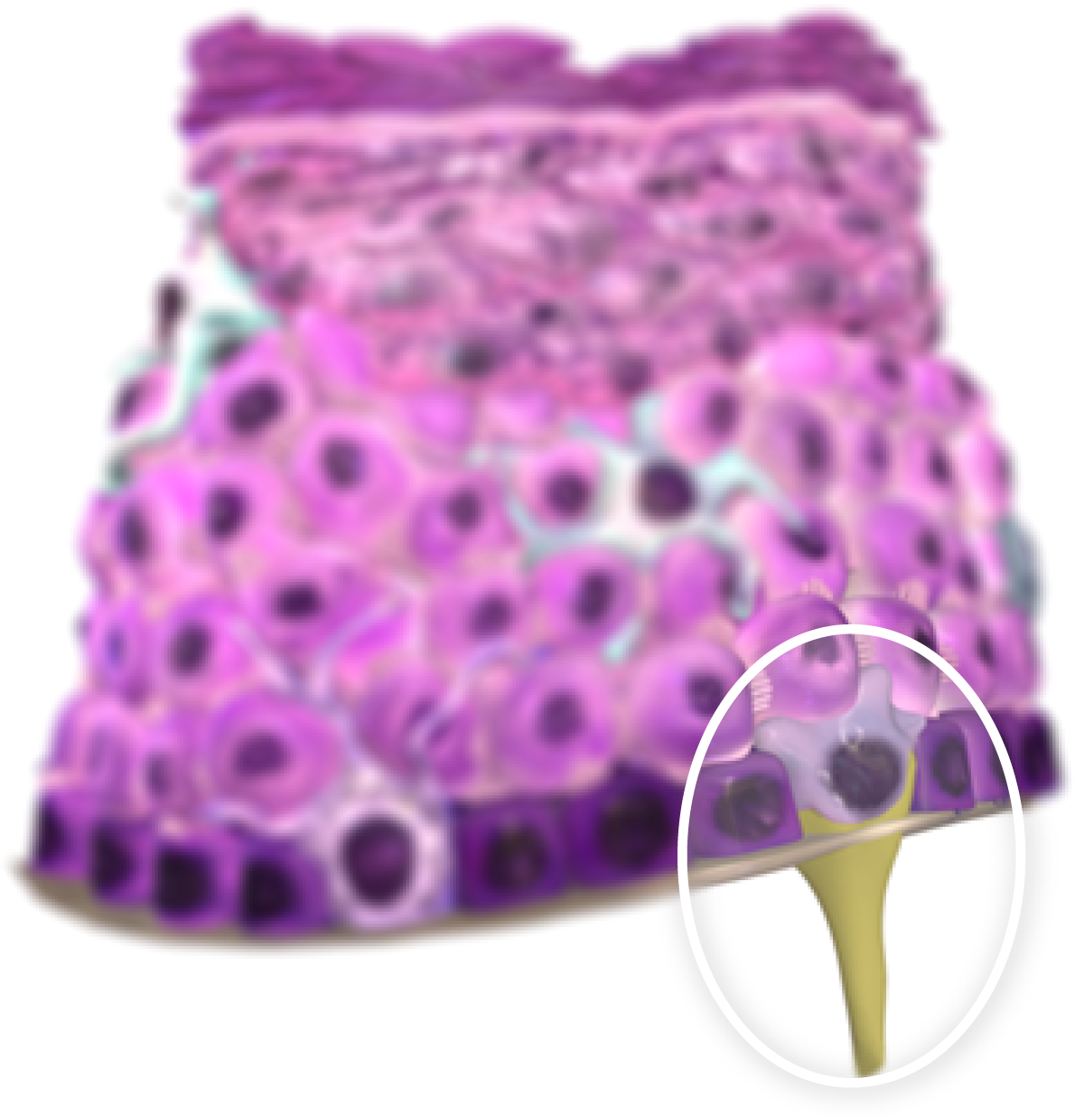
CELLS RESIDENT IN THE EPIDERMIS: MERKEL CELLS
A third cell type, the Merkel cell, is a specialized sensory transducer positioned amongst the cells of the basal epidermal layer. Although uncommon in most areas of the skin, they are often present in the skin of the digits and the lips and around hair follicles. Merkel cells are in communication with afferent nerve endings and are thought to modify the stimulus received by the sensory neurons.
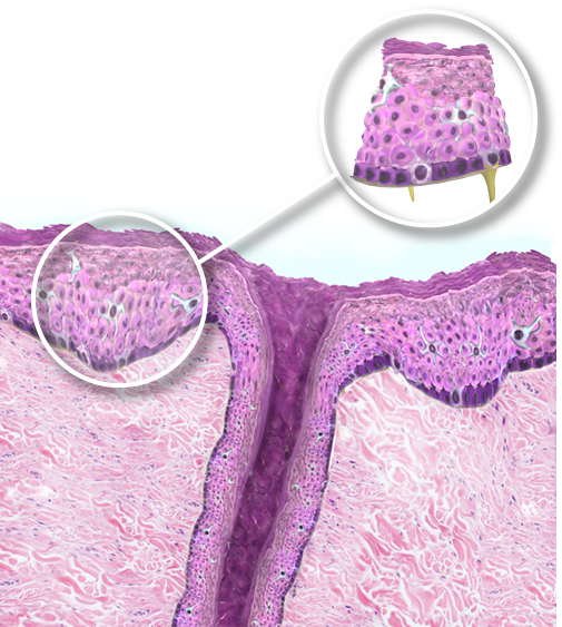
SURROUNDING STRUCTURES
In the image on the right the structures surrounding the model can be viewed, as well as the situation of the epithelium in relation tot them.
CLINICAL RELEVANCE: PEMPHIGUS
Pemphigus is one of a range of blistering conditions of the skin. It is an autoimmune disorder where the bodies own immune system is directed at the junctions that hold one squamous epithelial cell to another (desmosomes etc.). After being attacked the squamous cells are no longer anchored to each other and as a result blisters can form.
TUMOUR INVASION OF SURROUNDING STRUCTURES
Tumours can develop from any type of cell present within a tissue. In the skin, most commonly tumours arise from the epithelium.
BASAL CELL CARCINOMA
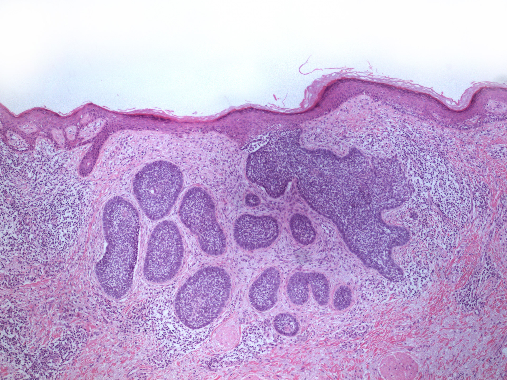
The image on the right shows buds and nests of tumour in the superficial dermis. These tumour nests are more darker staining than the very pink squamous epithelium of the overlying epidermis. The normal basal cells of the epidermis are also darker which is why these tumours are named basal cell carcinoma but it is not actually known what the primary cell of origin for these tumour is.
MELANOMA
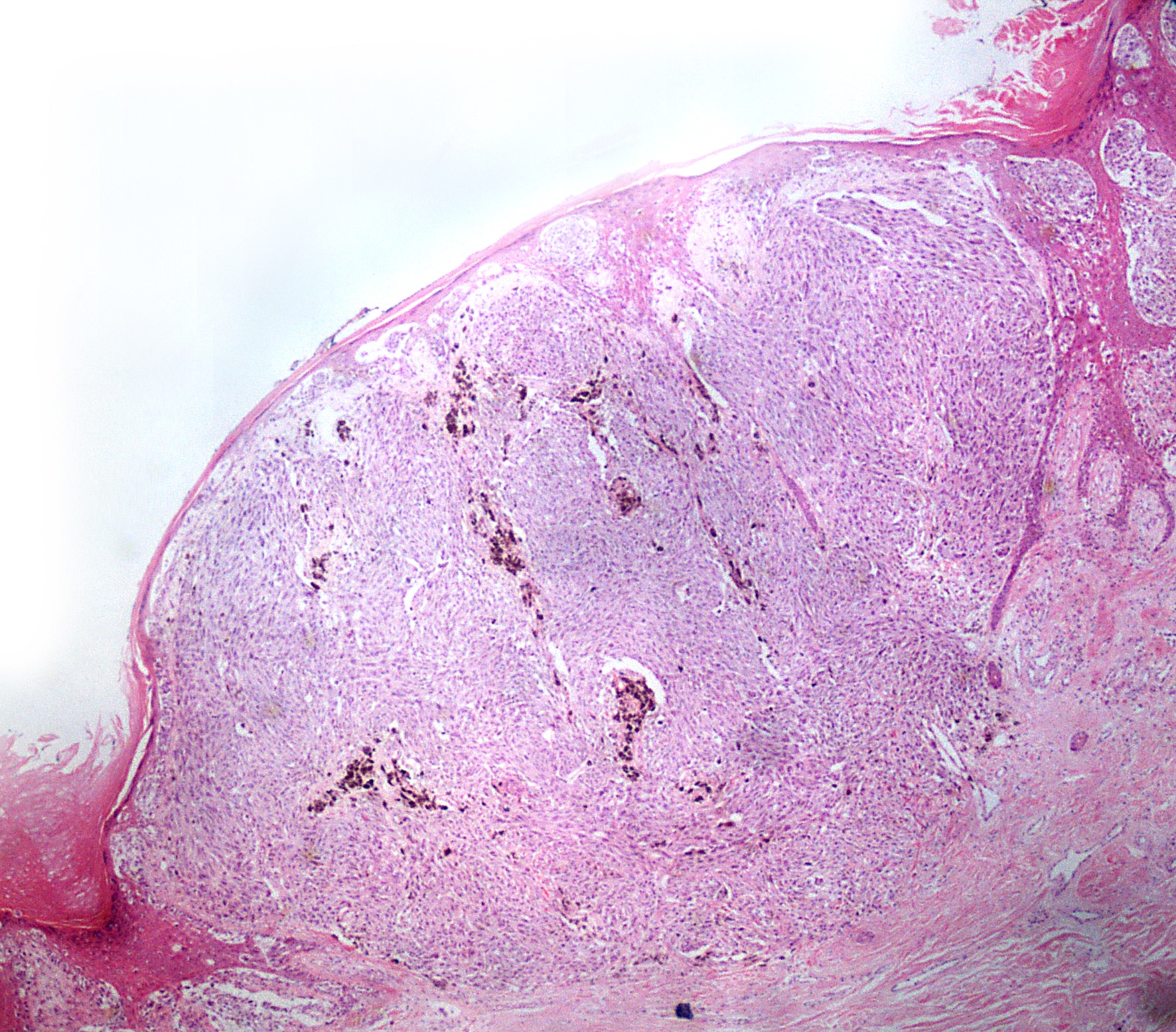
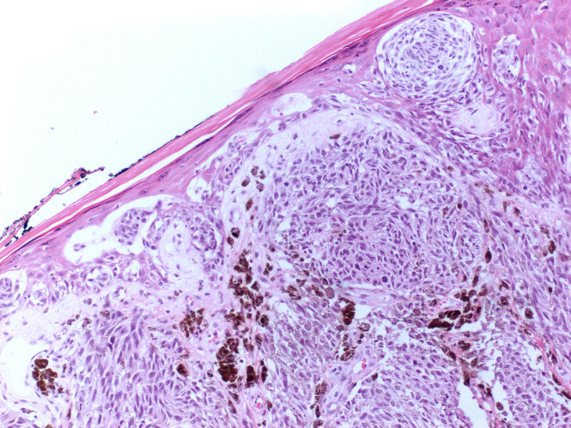
These two images below show a cutaneous melanoma in what is known as a vertical growth phase. Not all melanomas in the body produce melanin pigment but within the tumour shown here brown pigment can be easily identified. This irregular production of pigment gives rise to the asymmetry and colour variation that can be seen when examining abnormal moles with the naked eye.
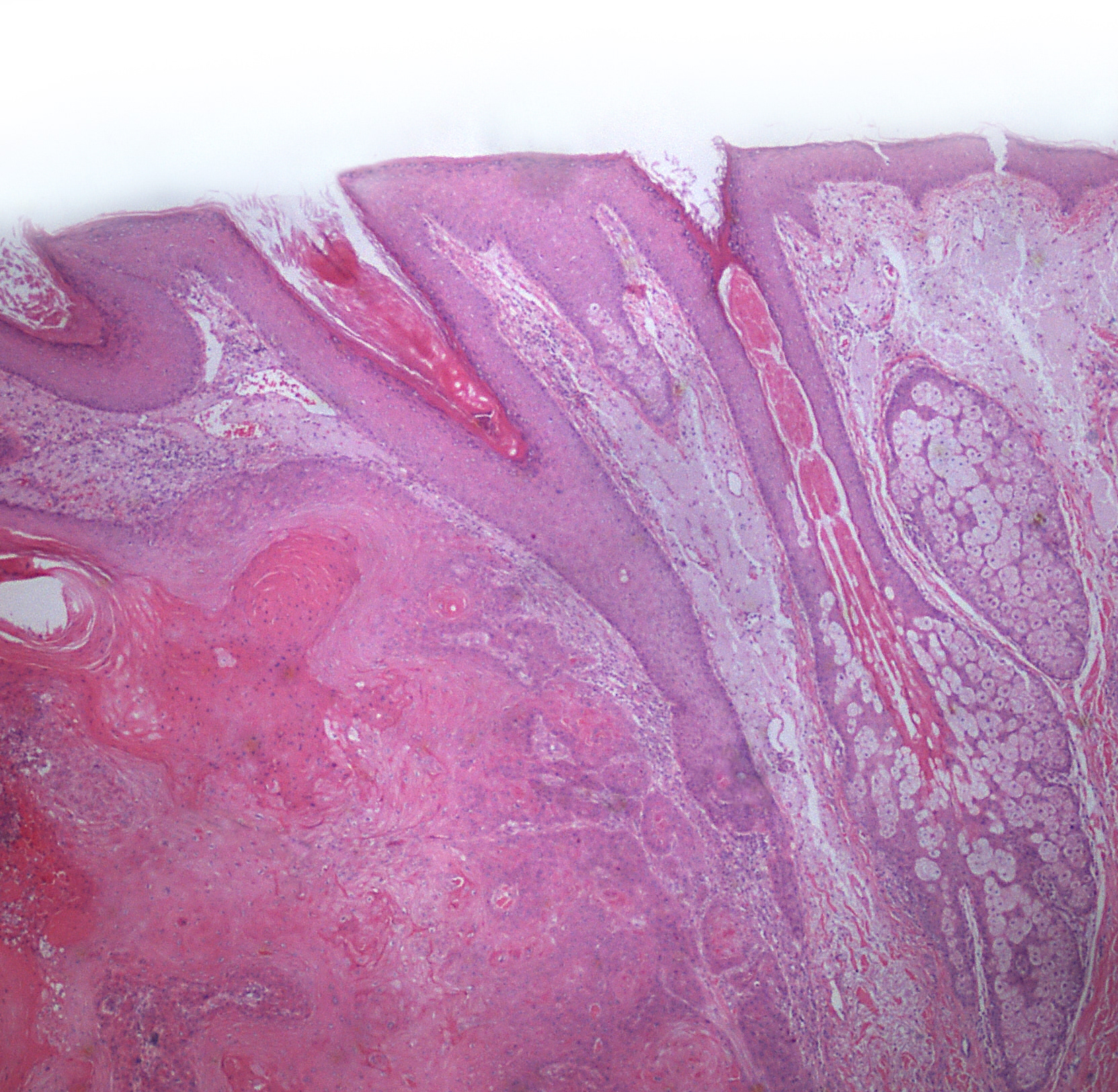
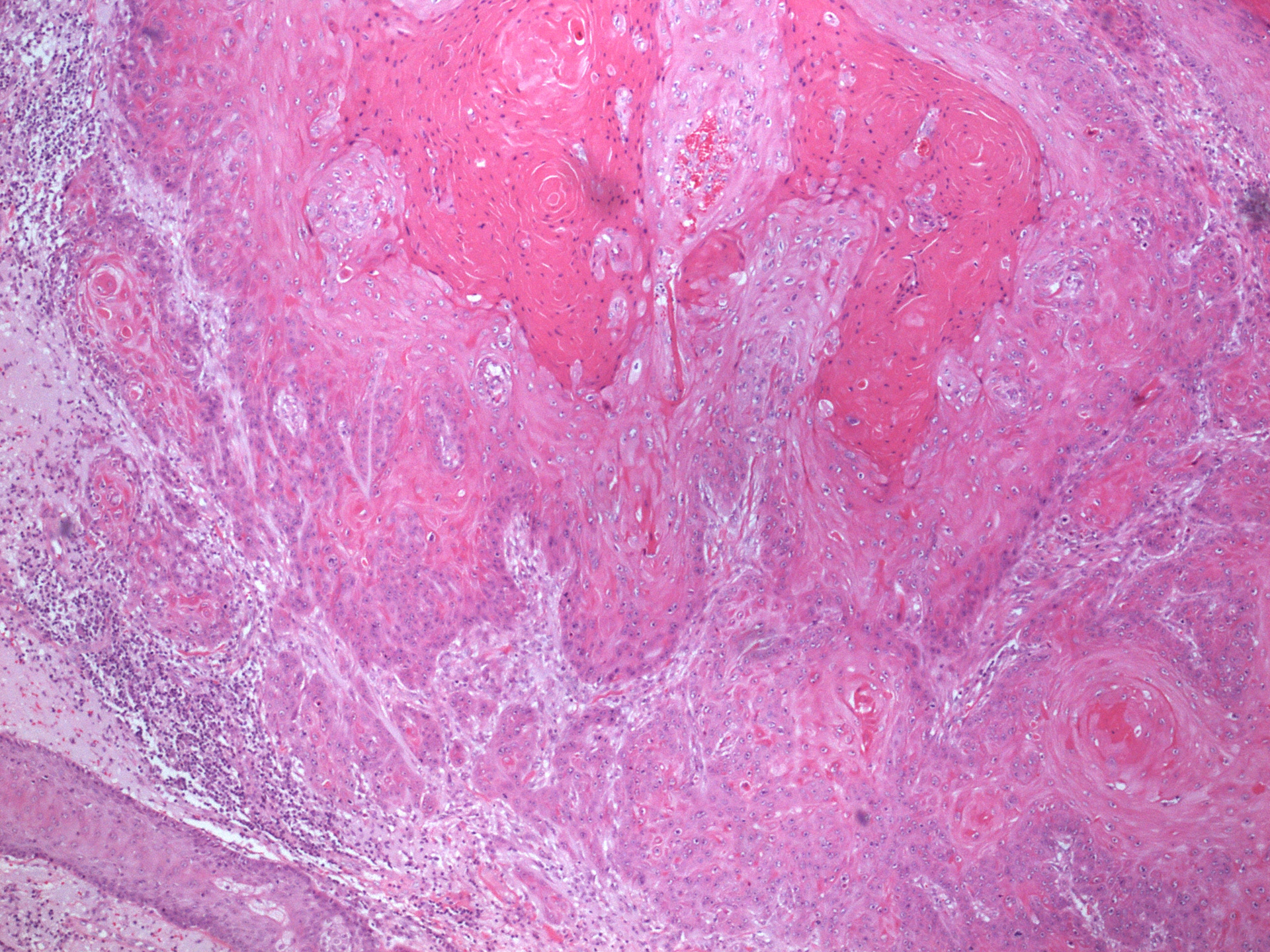
SQUAMOUS CELL CARCINOMA
One of the two images below is a low magnification view of skin. On the right of the image large hair follicles and their associated sebaceous glands can be seen. The sebaceous glands are made of large clear cells. On the left of this image you can see an infiltrating squamous cell carcinoma. Note the marked difference in colour and tone in contrast with the basal cell carcinoma. The higher magnification image shows keratin production by the tumour - proving this is a squamous cell carcinoma. The keratin is identified as much denser and deeply pink material towards the top right of the image.
SEE FOR YOURSELF
Want to have a look at the world through a microscope? Log in to the virtual microscope. Use your University of Dundee login information and have a look at a collection of microscopy images of different kinds of epithelium.